Radiology
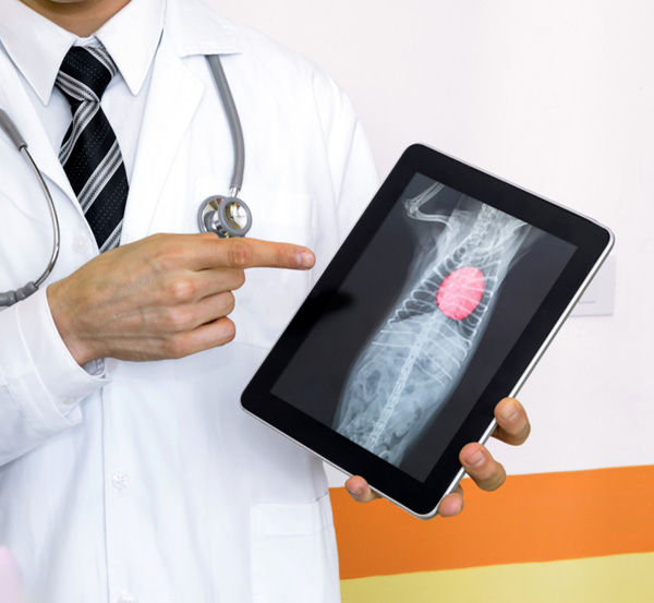
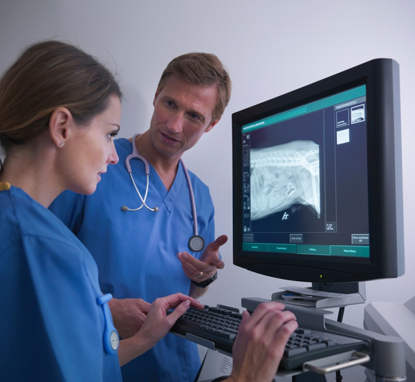
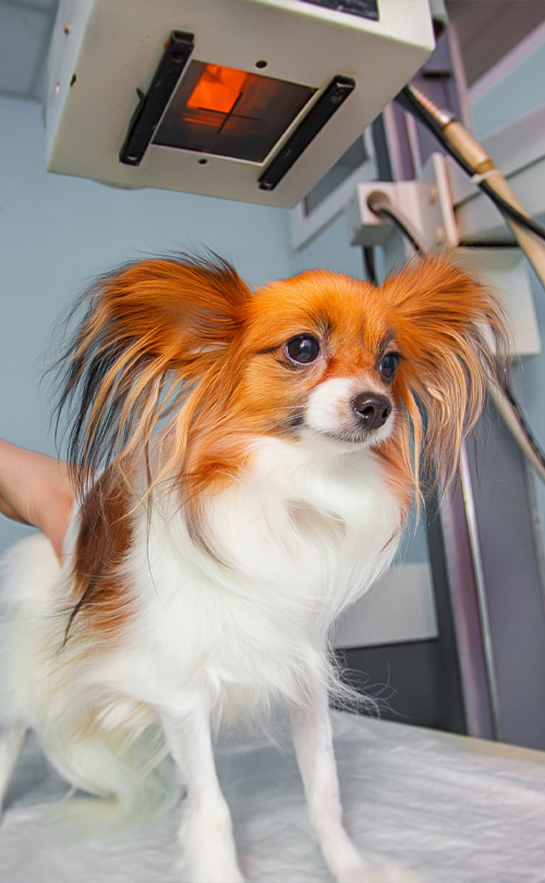
Veterinary radiology is a specialized field that uses noninvasive techniques and advanced imaging technologies to “see inside” the body in order to diagnose and help treat various medical conditions in animals. It is a central pillar of a veterinary hospital because it provides critical insights that guide the clinical decisions of all specialty and emergency services for most patients. From identifying fractures and tumors to assessing organ health, radiology plays a vital role in comprehensive pet care. Having an on-site, board-certified radiologist ensures that high-quality imaging and expert interpretation are readily available. We ensure every modality is used to its full diagnostic potential, elevating the standard of care and ensuring the best outcomes for our beloved animal companions.

Ultrasound (US)
Ultrasound uses sound waves that bounce off organs and tissues; these echoes are turned into real-time images showing the shape and movement of internal structures. It’s ideal for:
- Abdominal organ evaluation (liver, kidneys, spleen, etc)
- Detecting masses or free fluid
- Staging cancer
- Ruling out mechanical obstructions in the intestines or biliary system
- Diagnosing musculotendinous pathologies
Ultrasound allows us to see movement, like blood flow or bowel motility, and often guides minimally invasive procedures like needle aspirates or biopsies.
Coming Soon: Fluoroscopy
Fluoroscopy is real-time X-ray imaging. It will soon allow us to:
- Diagnose swallowing disorders
- Evaluate dynamic tracheal and bronchial collapse
- Characterize vascular anomalies
- Perform minimally invasive interventional procedures


